You don't need a chemistry background to compete, but it helps to understand the basics of how mass spectra are generated in a mass spectrometer:
Ionization: The instrument can only detect and measure charged ions, so first a molecule is ionized, picking up or losing charged species. In the instrument’s positive ion mode (more common for small molecules), the resulting ion is positively charged, in negative ion mode (less common but still used), it is negatively charged. The kind of charged ion the molecule acquires is called an adduct. For some common examples, the molecule may acquire a proton (positive), acquire an ammonium ion (positive), or lose a proton (negative). We would write these adduct forms respectively as [M+H]+, [M+NH4]+, or [M-H]-.
Precursor mass detection: The instrument measures the ion's precursor m/z (mass-to-charge ratio) with high accuracy, which strongly constrains the molecular formula. For small molecules, the charge (z) is usually +1 or -1, so m/z corresponds to the mass of the ion, and sometimes we will refer to m/z simply as “mass”.
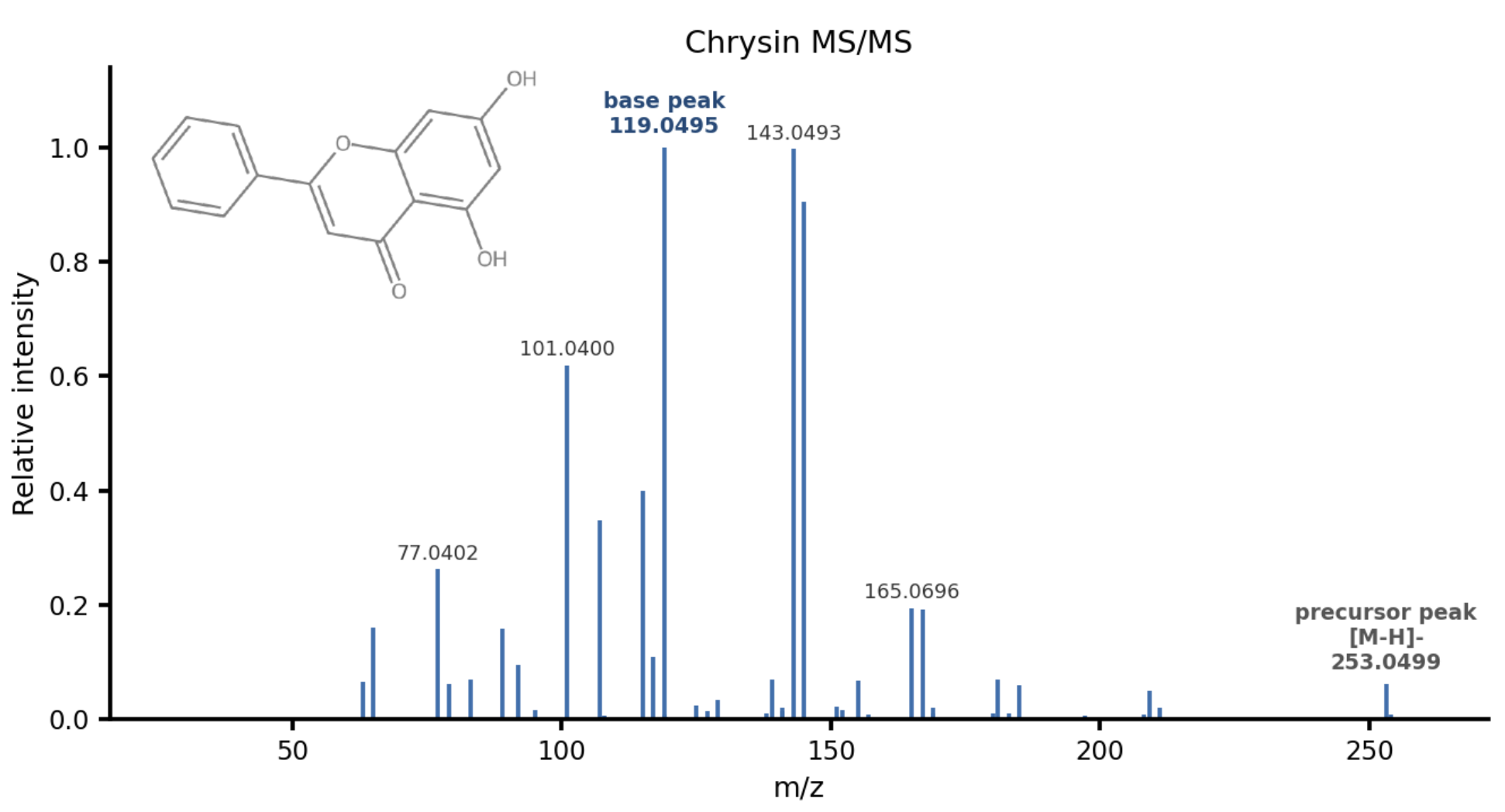
Fragmentation: The precursor ion (actually many individual ions of the same molecule+adduct) is then selected and fragmented by collision with neutral gas at a given collision energy, and the instrument records the m/z and intensity (abundance) of each fragment. The histogram of intensities across all detected fragments is the molecule’s mass spectrum. Intensities are typically normalized. A mass spectrum looks like this:
Figure 1: MS/MS spectrum of Chrysin [M+H]- adduct at around 50eV, tims(Source: Enveda).
Each m/z is called a fragment ion or peak. The base peak is the highest-intensity fragment ion. The precursor peak is the m/z corresponding to the intact, unfragmented ion. It may or may not be present, depending on how thoroughly the ion was fragmented.